Proptosis
Proptosis is defined, as the forward displacement of the globe (eyeball) out of the socket, with the eyelids trapped behind the globe.
Proptosis is an ophthalmic emergency. Any suspected trauma to your pet’s eye warrants a visit to your veterinarian immediately.
Let us first have a look at the normal eye anatomy:

Predisposing factors: Breed predisposition
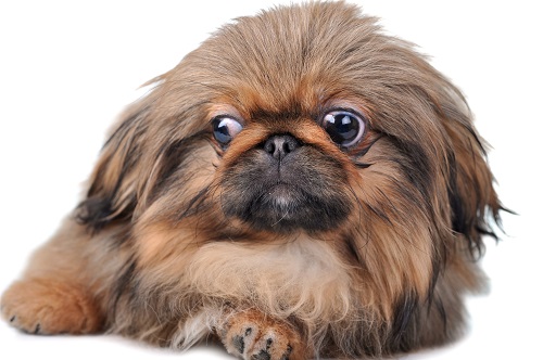
Proptosis is a condition more commonly seen in Brachycephalic breeds (dogs with prominent bulging eyes, short noses and shallow eye sockets). Pekingese, Pug, Boston terrier and Shihtzu are over represented.
Common Causes
- Trauma is the most common cause of proptosis. In small breed dogs this often occurs during a fight with a larger dog, wherein the larger dog bites over the scruff on the neck and starts shaking the smaller dog. The pulling of the skin back over the head allows for the eye to pop forward, out of the socket. Bite wounds directly to the face could also result in proptosis of one or both eyes but this is very rare.
- Blunt trauma to the face or head, often seen when hit by a car.
- Brachycephalic breeds have very shallow eye sockets, and very large openings of the eyelids, in these breeds even mild manual restraint can result in proptosis.
- Space occupying lesions behind the globe e.g. tumours located behind the eyeball, applying pressure and pushing the globe forward.
Clinical signs
Often the first thing noticed by owners is a very prominent eye where the eyelids are unable to blink over the eyeball (globe).
Other signs include:
- Swelling and inflammation of the tissue/muscles surrounding the proptosed globe
- Bleeding inside the eye
- Abnormal pupil (it may be dilated or constricted)
- Rupture of the globe
- Rupture of the muscles around the globe, the eye then deviates outward. These muscles are responsible for keeping the globe in place and are responsible for movement of the eye.
Proptosis needs to be distinguished from
- Bupthalmia: Enlargement of the eyeball (globe), often caused by Glaucoma or a tumour in the eye.
- Exopthalmia: Abnormal protrusion of the globe forward, the size of the eyeball remains normal it is just the positioning that changes. Often caused by tumours behind the globe, abcesses in the tissues surrounding the eye or bleeding behind the globe.
What to do when you notice your pet’s eye has proptosed?
- Keep the proptosed eye moist. You can use lubricating human eye drops, or sterile water, try not to use tap water as tap water contains micro-organisms which can be detrimental to eye health.
- If you have an old Elizabethan collar you should put it on your dog as a proptosed eye is very painfull and your dog will try to rub it or scratch at it using its paw. The Elizabethan collar will therefore aid in limiting further trauma to the proptosed eye.
- Do not give your pet any human pain medication or anti-inflammatories, your vet will treat it as soon as it arrives at the practice with the correct medication at the correct dosage.
 Presentation to the vet
Your pet will be assessed for any other injuries or possible complications; this is a very important step especially if the cause of the proptosis was an accidental hit by car. The patient must be stable before any surgery can be attempted.
The vet will then examine both eyes to assess the extent of injury.
The vet will look at the cornea (thin see-through layer) by staining it with fourecein to determine if there are any cuts or scratches on the surface of the eye (this stain changes color from orange to green if there are any cuts or scratches on the cornea). Then also the muscles, skin and nerves (optic nerve) attached to the eye will be examined
Based on the severity of the injury two treatment options exist, either replacement of the eye back into the socket or enucleation (surgical removal of the proptosed eye).
The prognosis for vision in the affected eye is always poor and is often dependant on the extent of the trauma to the eye and how soon treatment is started. Even if vision is lost, rapid response could aid in salvaging the eye for cosmetic reasons.
When the patient is stable enough they are placed under general anesthesia.
If the eye can be salvaged it is lubricated and placed back into the eye socket. Sometimes an incision is made on the outer edge of the eyelid to allow more space and make replacement of the eye easier. They eyelids are then stitched closed – this procedure is called a temporary tarsorrhaphy. Often times the vet will dispense eye drops that needs to be applied daily, this promotes healing of the cornea. The stitches are removed after two weeks and the function of the eye is reassessed to determine if the vision in the eye was affected or not.
If an enucleation is required, the globe is removed, blood vessels and nerves are tied-off and excessive tissue is also trimmed away. The eyelids are permanently stitched closed.
In both instances the patient is sent home with pain medication and an Elizabethan collar to prevent further trauma to the surgical site.
The thought of imagining a beloved pet with only one eye is very distressing and traumatic to owners, but most often these patients recover really quickly and do lead normal happy lives.
© 2018 Vetwebsites – The Code Company Trading (Pty.) Ltd.